 |
 |
 |
 |
 |
||||||||||||
|
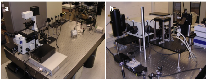
Steve Smith — NanoScience and NanoEngineering Nano-fs — Nanophotonics Laboratory: 660 Sq. Ft. laser laboratory with chilled water, clean power and automated backup battery supply. Appended 8'x16' wet bench, overhead shelving, chemical storage cabinetry and bacterial cell culture tools. 3 optical tables equipped with state of the art continuous wave and femtosecond pulsed laser systems integrated into custom built imaging systems as described below: Single Molecule, Superresolution Energy and Time-resolved Imaging PALM/TIRF system: A custom built TIRF system, based on an Olympus IX71 inverted frame microscope, equipped with a C-Lock focusing servo system and a back illuminated CCD, located on a 4'x6' optical table is devoted to this activity and is equipped for PALM and DOPI imaging. The system includes computer controlled shutters, visible and UV laser sources and is controlled through LabView software (figure a). Energy- and Time-Resolved Photoluminescence Imaging: This custom built spectral imaging system includes an HR640 single grating spectrometer (high dispersion gratings), Princeton Instruments SPEC-10 back illuminated CCD, Millenia 5W pump laser and KML Ti:Sapphire femto-second laser, and a closed-loop piezo- electric stage. A time-correlated single photon counting card allows lifetime measurements. MPD fast avalanche photodiode and Multi-channel scaler allows low rep rate laser single photon counting. Housed on a 4'x8' optical table (Figure b)
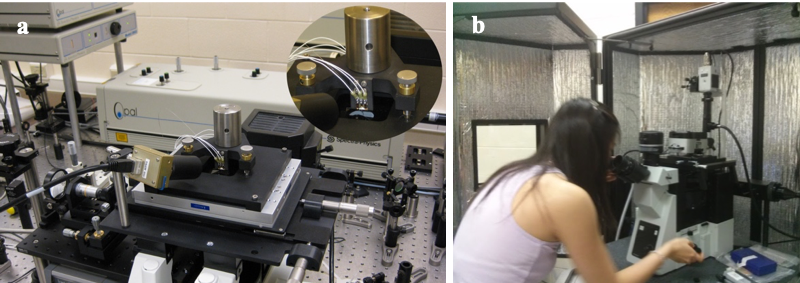
Multiphoton and Correlative Fluorescence / AFM Imaging Multiphoton near-field / wide-field scanning imaging system: Computer controlled piezoelectric stage (MadCity Labs), Olympus IX71 inverted frame fluorescence microscope with EMCCD (Andor iXon 512). The system operates as a near-field / widefield spectrally resolved multi-photon imaging system. The optical sources are a Millenia Pro 10W pump laser, Tsunami Ti-Sapphire femtosecond Oscillator, an OPAL Optical Parametric Oscillator (tunable from 550nm to 2.4μm, by frequency doubling), and miscellaneous low power lasers (532nm-633nm). The system housed on a 4x8 optical table (Figure a). Combined AFM and Fluorescence Microscopy: (in adjacent 150 sq. ft. lab). An Asylum MFP-3D atomic force microscopy is integrated with an Olympus IX81 inverted fluorescence microscope equipped with a scientific CMOS camera (ORCA FLASH 4.0 sCMOS), integrated with custom built photo-activated light microscopy (PALM) system with 561nm and 405nm excitation and activation wavelengths (Figure b).
Energy and Time-resolved Spectroscopy Ultrafast Femtosecond Spectroscopy: The optical sources available are a Millenia Pro 10W pump laser, Tsunami Ti-Sapphire high-power femtosecond Oscillator, and an OPAL tunable Optical Parametric Oscillator (tunable from 550nm to 2.4μm, through idler and frequency doubling), and miscellaneous low power lasers (532nm-633nm) and associated optics and electronics. Optical Spectroscopy: Perkin-Elmer Lambda-650 UV/Vis spectrometer, a Perkin-Elmer LS-55 fluorescence spectrometer, and a home built photoluminescence spectroscopic setup equipped with a liquid Helium bath-style cryostat (Oxford OptistatSXM) for variable temperature studies.
| ||||||||||||||||